viernes, 30 de octubre de 2015

cancer

 
Prostate cancer:

The prostate produces a fluid that forms part of semen. The prostate is located just below the bladder, in front of the rectum. It surrounds the urethra (the tube that carries urine and semen through the penis and out of the body).

Almost all prostate cancers are adenocarcinomas (cancers that begin in the cells that release mucus and other fluids). Prostate cancer often has no early symptoms. Advanced prostate cancer may make men more frequently urinate or have a weak flow of urine, but these symptoms are also due to benign prostate conditions.


Prostate cancer usually grows very slowly. Most men with prostate cancer are over 65 and do not die from the disease.


You can use the following tests and procedures:

Physical exam and history: An exam of the body to check the overall health and identify any signs of disease, such as lumps or anything else that does not look normal. Data on the patient's health habits and history of illnesses and treatments in each case are also taken.

Digital rectal exam (DRE): An exam of the rectum. The doctor or nurse inserts a finger in a lubricated finger into the rectum and feels the prostate through the rectal wall for lumps or abnormal areas.


Anatomía del sistema reproductor y el sistema urinario masculino; el dibujo muestra vistas frontales y laterales de los uréteres, los ganglios linfáticos, la vejiga, el recto, la glándula prostática, la uretra, el conducto deferente,  el pene y los testículos, la vesícula seminal, y el conducto eyaculatorio.Un dibujo en dos paneles muestra la anatomía reproductora y urinaria normal así como hiperplasia prostática benigna (HPB).  El panel de la izquierda muestra la próstata normal y el flujo de orina de la vejiga a la uretra.  El panel de la derecha muestra un agrandamiento de próstata que ejerce presión sobre la vejiga y la uretra, con la obstrucción del flujo de la orina.


opinon: This cancer is very aggressive for men, and is more common in men over 65, it is recommended that a review be conducted at least 2 times a year.


bibliography: 

http://www.cancer.gov/espanol/tipos/prostata/paciente/tratamiento-prostata-pdq

Instituto Nacional Del Cáncer


CRANIOTOMY

A craniotomy is a surgery in which part of the skull bone is removed in order to expose the brain and the structures of the central nervous system, the bone flap is temporarily removed and the end of the surgery is repositioned to give new protection the brain and its structures.

It is important to differentiate it from the craniectomy, which is a similar procedure that involves the permanent removal of the skull, in order to make room for the brain when there is significant inflammation.


A craniotomy may be performed for different reasons, such as to treat the following diseases:

Resection of brain tumors
Repair of brain aneurysms
Removing both subdural, epidural or intracerebral hematomas.
Treatment of congenital arteriovenous malformations
Brain abscess drainage
Repair skull fractures secondary to trauma
Repair of the membranes covering the brain (dura) when there
Cerebrospinal fluid leak.

A craniotomy may be performed in any of the skull and is named according to the part of the bone is resected, so craniotomy may involve one, two or even three combination skull bones and calling for example, frontal craniotomy if is only the frontal bone, frontotemporal craniotomy if the front part of the temporal bone and which is resected or excised if frontoparietotemporal craniotomy of the frontal, parietal and temporal bone.

The procedure is performed with the patient completely asleep with general anesthesia. The position depends on the site where it will perform a craniotomy, can be placed face up, face down, sideways, his head turned to one side or another etc. in any case, the fixation of the head is necessary to prevent movement during surgery.

Decontamination of the surgical site with bactericidal substances is performed and proceeds with aseptic technique to incision of the scalp and may be linear, curved or horseshoe provided in area where hair grows, for cosmetic issues


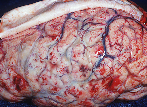
Craniotome a hole using a bone from which extends a wide enough window of bone (craniotomy) to adequately treat any injury is done. Immediately below are bone coverage of the brain called the meninges, the outermost of which the dura and the rest must be opened to perform the required treatment for each patient

As with any surgical procedure, complications may arise, among which include, but are not limited to:

• nervous system infection or wound
• hemorrhage (bleeding)
• thrombi (blood clots)
• CSF leak
• damage to blood vessels that may condition infarcts
• slurred speech, strength, mobility and coordination of the limbs.
• Alterations to walk
• death


In my personal opinion, I can say that this surgery is one of the most interesting in the field of neurology as it involves many immersed procedures into one, for me is the most beautiful because it takes many hours of surgery and requires a lot accuracy to conclude successfully

Recovered of http://www.cirugia-neurologica.org/cirugia-cerebral.ws on 30th October,2015 at 15:00
Rebeca L. Guzmán L.


lunes, 26 de octubre de 2015


NEONATAL MENINGITIS


Is defined as neonatal meningitis clinical situation characterized by signs and symptoms of systemic infection, inflammatory markers compatible (blood count, C-reactive protein (CRP), etc.) and changes in the cerebrospinal fluid (CSF) suggestive of meningeal inflammation (increased leukocyte and protein and decreased glucose) isolation of microorganisms not be indispensable. For the interpretation of the findings in the CSF should be borne in mind that in the neonatal period normally ranges are different to those found in infants and children, and also differences between newborns (RN) and term neonates.
Once the diagnosis is made and based on the results of microbiological analysis it is considered microbiologically proven meningitis when CSF culture positive for bacteria, viruses or fungi; microbiologically likely meningitis when CSF culture is negative and positive blood culture and meningitis microbiologically unproven when both cultures are negative. It is unclear why in the course of a few RN bacteremia develop meningitis and some not. Experimental studies postulate that the high number of bacteria that invade the bloodstream correlates with the likelihood of meningitis. It also relates to the characteristics of germs, and thus the capsular antigen of E. coli K1, which is similar to the capsular polysaccharide of N. meningitidis type B, it is important to promote the survival of the organism in the bloodstream and promote their step by the blood-brain barrier. Over 80% of neonatal meningitis E. sliding block caused by strains carrying this antigen. Similarly was separated ñalado in infections with S. agalactiae (GBS), an increased risk of developing meningitis when the infection is by biotypes B1 to B6 or importance of the type III capsular polysaccharide as a virulence factor in Late meningitis caused by this germ.

VERTICAL MENINGITIS


They are caused by germs initially localized in the maternal genital channel, via ascending or direct contact, first pollute the skin and / or respiratory mucosa and / or digestive fetus and then invade the bloodstream. MTCT risk factors obstetric those circumstances that are associated with a significant incidence of infection bacteriana6 considered: maternal vaginal colonization by pathogen, spontaneous preterm birth, chorioamnionitis, intrapartum fever, prolonged rupture of membranes, hypoxia, urinary tract infection at the end of the gestation, etc.


NOSOCOMIAL MENINGITIS

They are caused by germs located in the hospital environment (especially in the neonatal ICU) that first colonize the skin and / or mucous membranes, then through the cutaneous and mucosal barrier and invade the bloodstream and ultimately cross the blood barrier -encefálica and lead to meningitis. Risk factors are considered: enter the NICU, premature, after broad-spectrum antibiotics, use of central catheters to infuse intravenous feeding, intubation and mechanical ventilation, and in case of meningitis also matter congenital anomalies (meningocele, myelomeningocele, sinus dermicus, etc.) that favor the direct invasion of bacteria into the subarachnoid space and neurosurgical techniques (ventricular puncture, placement of reservoir placement of ventricular-peritoneal drainage, intracranial surgery, etc.) that favor the direct implant of bacteria in the central nervous system.


OPINION

On the subject of meningitis is a broad topic as it affects infants , children and adults what a vast subject , in this section I covered the topic of meningocele in neonates.
When reading about it , I could percatar neonatal meningitis in two types which are vertically and nosocomial . Which trigger other diseases in the neonate which result in more complications for the newborn.

BIBLIOGRAPHY
Meningitis neonatal

B. Fernández Colomer, J. López Sastre, G. D. Coto Cotallo,
A. Ramos Aparicio, A. Ibáñez Fernández.
Servicio de Neonatología
Hospital Universitario Central de Asturias. Oviedo


https://www.aeped.es/sites/default/files/documentos/22_0.pdf

sábado, 24 de octubre de 2015

Revista chilena de pediatría - Falla cardíaca como manifestación temprana de hipertiroidismo neonatal: Caso clínico

girls I share an article about a case of newborn with hyperthyroidism and heart failure, if I would like to read it and they think if we do this activity more dynamic and add me as a comment under their own assessment and judgment a nursing diagnosis that create identify the patient's needs.


CLICK AQUI  -->Revista chilena de pediatría - Falla cardíaca como manifestación temprana de hipertiroidismo neonatal: Caso clínico


atte JSH

miércoles, 21 de octubre de 2015

Anatomy

The central nervous system (CNS) consists of the brain and spinal cord. All of it is protected by a covering of bones:

·         The brain by the skull
·         Spinal cord spine

Between the bone and brain or spinal cord, other protective layers of connective tissue both in the region of the brain and spinal cord are called meninges, which are formed by three types of membranes (dura, are arachnoid and pia mater). These membranes are distinguished in two types:

·         The dura (outer and meninges attached to the bone structure)
·         The leptomeninges, which is inside

The leptomeninges is composed of two membranes, in turn, interconnected:

·         Arachnoid
·         The pia mater directly adhered to the CNS and the circulating blood vessels


Arachnoid tissue has no blood vessels and is above fibers connected with vascularized connective tissue pia mater. Between the arachnoid and pia mater is the subarachnoid space, through which the cerebrospinal fluid (CSF).


MENINGES

CNS (brain and spinal cord) is surrounded by three layers of tissue called meninges. There are three meningeal layers:

1. Dura: the outermost layer and stronger. It consists of tissue
irregular dense connective. It is attached to the bone. Presents projections shaped partitions that separate areas of the brain:
2. Hoz brain: a vertical partition and medium located between the two cerebral hemispheres in the interhemispheric fissure.
3. Tentorio or tentorium: is situated perpendicular to the sickle, separating the brain structures of the posterior fossa (brain stem and cerebellum).
4. arachnoid: below the dura. It consists of tissue avascular connective rich in collagen and elastic fibers that form as a
mesh. Between the meninges and dura mater is the subdural space.
5. Pia mater: a very thin, transparent layer of connective tissue that is intimately attached to the central nervous system which covers. Between arachnoid and pia mater is the subarachnoid space, which contains cerebrospinal fluid.

OPINION

To better compreder a disease , it is important to know the anatomy of the body as it is very important to detect which part of the body is affected and that treatment is going to provide .

The anatomy of the human body is complex , but yet know is very important to understand that part of the body is affected and this will have complications with other devices and sitemas that make up the body .

BIBLIOGRAPHY:

·         http://www.hvn.es/servicios_asistenciales/medicinainterna/ficheros/meningitis20052006.pdf

cancer

What is breast cancer?

The breast or breast consists of fat, connective and glandular tissue. Each breast is between 10 and 20 sections known as lobes, which in turn are divided into smaller sections, the lobules. The lobules contain milk-producing glands during lactation. Through the milk ducts to the nipple arrives.


Lobules and ducts are in the stroma, adipose tissue in which the blood and lymph vessels, lymph nodes are also located. These nodes are responsible for protection against bacteria, tumor cells and other harmful substances.

Breast cancer occurs when cells of the glandular epithelium reproduce uncontrollably and very quickly. These cancer cells can travel through the blood and lymphatic vessels and reach other parts of the body, where they can adhere to the organs and form metastases.

Breast cancer can occur in both men and women. However, over 99 percent of diagnoses occur in women.


Types: 
  • ductal carcinoma
  • Lobular carcinoma or lobular
  • Inflammatory breast cancer 
  • Cancer Paget


bibliography: http://www.dmedicina.com/enfermedades/cancer/cancer-mama.html


opinion: Breast cancer is a malignant disease for women because they can ralizarse as a mastectomy procedure to remove the mammary glands (may be both or one) and in this case men also precent; but less incidensia because breast cancer is associated more females.

public: V. montserrat, H.C.


sábado, 17 de octubre de 2015

labor and vaginal childbirth

This video shows labor and vaginal birth. It includes the three stages of labor: dilatation and in Its active and latent phases, the expulsion When your baby exits the birth canal, and finally delivery of the placenta.
 I invite you to observe in a very interactive and easy to understand video.

JSH

cancer

Cancer of the cervix or cervical cancer

It is the abnormal growth of cells found in the cervix. Initially, the lesions are so small they can not be seen with the naked eye and thus last several years.

When the cancer is in an advanced stage you can be seen with the naked eye in the gynecological examination or cause other troubles, including abnormal bleeding after intercourse, between periods or after periods of menopause, increasing the flow of bleeding smelly genital tract, hip pain and weight loss.

When you first started, the treatment can be surgically removing the matrix or with radiation therapy and chemotherapy in advanced cases.


Women more likely to have this cancer are those that:


  • They started their sex before age 18
  • They have had more than 3 sexual partners
  • They have had more than 3 deliveries
  • smoke
  • They have problems of malnutrition
  • They are infected with HPV in the cervix


IMSS
bibliagraphy: http://www.imss.gob.mx/salud-en-linea/cancer-cervico-uterino


comment: This type of cancer is very severe and very evil for women, since it is a disease with a very high incidence among the Mexican population; prebenible when the disease is detected early, so women should perform a checkup at least every 3-6 months.

public: valeria M.H.C


viernes, 16 de octubre de 2015

Bone Marrow Aspiration and Biopsy


OPINION
The video is teaches us how to learn about bacterial meningitis vaccines and the global need for improved strategies for the prevention of meningococcal disease.
It's a short but very clear and understandable video.

BIBLIOGRAPHY

https://www.youtube.com/watch?v=LjxmoIwxolU
TYPES OF MENINGITIS

Aseptic meningitis


It is usually benign course and in most cases aetiology viral. There may be non-infectious causes of aseptic meningitis. 

Has a biphasic clinical course. Preceding his first appearance of symptoms or signs of respiratory, gastrointestinal or viral skin condition, occurring meningeal later stage with more specific symptoms such as headache, fever and meningeal signs. Symptoms are uncommon commitment brain such as seizures or altered mental status.


Septic meningitis (bacterial meningitis)
The bacterial meningitis on the other hand imply greater morbidity and mortality and require rapid diagnosis and treatment to avoid a fatal outcome or irreversible consequences therefore is a neurological emergency. The overall rate mortality is 25 % and up to 60 % morbidity.


OPINION
My opinion on the issue is to speak of meningitis is very extensive , as there are specific types of the disease, and each has multiple afectacines in orgaismo of those who suffer , I consider it important to know the types of meningitis, for identification in affected patients and also see that he will provide treatment.

BIBLIOGRAPHY:
http://www.acnweb.org/guia/g1c10i.pdf


domingo, 11 de octubre de 2015

What happens when your DNA is damaged? - Monica Menesini


partners, the topic of genetic mutations explains the types of errors that occur during DNA replication, as well as the agents that cause mutations, leave you a video that explains how an error in genes is repaired, a video very simple, but it's a topic they already know


cancer

most common types of cancer:

The list of common cancers including cancers that are diagnosed more frequently in the United States (the non-melanoma skin cancers are not included):

Bladder cancer
Breast cancer (breast)
Colon and rectum
Endometrial cancer
Kidney cancer (renal cell)
Leukemia
Lung cancer
Melanoma
NHL
Pancreatic cancer
Prostate cancer
Thyroid cancer

For a cancer is considered common, the estimated number of new cases for 2015 had to be 40,000 or more.

The most common type of cancer in the list is breast cancer, with over 234,000 new cases estimated in the United States by 2015. The next most common cancers are prostate cancer and lung cancer.

Because the colon and rectum are often called "colorectal cancers," both types are combined in the list. In 2015, the estimated number of new cases of colorectal cancer is 93 090 and 39 610, respectively, for a total of 132 700 new cases of colorectal cancer.


Instituto Nacional del Cáncer
de los Institutos Nacionales de la Salud de EE. UU.


bibliography: http://www.cancer.gov/espanol/tipos/comunes


opinion: in my opinion, there are several types of cancer, some are more aggressive than others, but the desert is a disease that is very common today, and is silent; since most precent no symptoms until already advanced the disease.


cancer

what is menaloma?

The most dangerous form of skin cancer, these cancerous growths develop when unrepaired DNA damage to skin cells (most often caused by ultraviolet radiation from sunshine or tanning beds) triggers mutations (genetic defects) that lead the skin cells to multiply rapidly and form malignant tumors. These tumors originate in the pigment-producing melanocytes in the basal layer of the epidermis. Melanomas often resemble moles; some develop from moles. The majority of melanomas are black or brown, but they can also be skin-colored, pink, red, purple, blue or white. Melanoma is caused mainly by intense, occasional UV exposure (frequently leading to sunburn), especially in those who are genetically predisposed to the disease. Melanoma kills an estimated 9,940 people in the US annually.

If melanoma is recognized and treated early, it is almost always curable, but if it is not, the cancer can advance and spread to other parts of the body, where it becomes hard to treat and can be fatal. While it is not the most common of the skin cancers, it causes the most deaths.
© 2015 The Skin Cancer Foundation
bibliography: http://www.skincancer.org/skin-cancer-information/melanoma

     Resultado de imagen para melanoma     

cancer

what is menaloma?

The most dangerous form of skin cancer, these cancerous growths develop when unrepaired DNA damage to skin cells (most often caused by ultraviolet radiation from sunshine or tanning beds) triggers mutations (genetic defects) that lead the skin cells to multiply rapidly and form malignant tumors. These tumors originate in the pigment-producing melanocytes in the basal layer of the epidermis. Melanomas often resemble moles; some develop from moles. The majority of melanomas are black or brown, but they can also be skin-colored, pink, red, purple, blue or white. Melanoma is caused mainly by intense, occasional UV exposure (frequently leading to sunburn), especially in those who are genetically predisposed to the disease. Melanoma kills an estimated 9,940 people in the US annually.

If melanoma is recognized and treated early, it is almost always curable, but if it is not, the cancer can advance and spread to other parts of the body, where it becomes hard to treat and can be fatal. While it is not the most common of the skin cancers, it causes the most deaths.
© 2015 The Skin Cancer Foundation
bibliography: http://www.skincancer.org/skin-cancer-information/melanoma

Resultado de imagen para melanoma     Resultado de imagen para melanoma